Understanding Arthrograms: Purpose, Procedure, and Potential Risks

Understanding Arthrograms: Purpose, Procedure, and Potential Risks
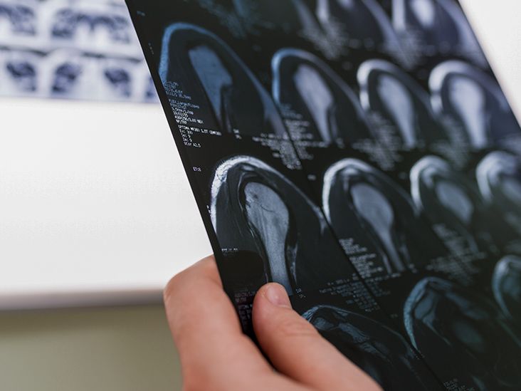
An arthrogram is a specialized imaging technique designed to provide a clearer view of your joints. This procedure involves the injection of a contrast medium, often referred to as dye, into the joint space. Following the injection, advanced imaging methods, such as X-rays, fluoroscopy, MRIs, or CT scans, are utilized to visualize the area in detail.
Arthrograms are instrumental in diagnosing issues that may contribute to joint pain or impaired function, allowing healthcare providers to discern finer details in tissues and bones. While generally safe, this test is typically contraindicated for individuals with joint infections, certain types of arthritis, or those who are pregnant. In the following sections, we will explore the various types of arthrograms, what to anticipate during the procedure, and the patient profiles that are considered good candidates for this test.
What is the Purpose of an Arthrogram?
An arthrogram is primarily employed to identify the underlying causes of joint discomfort or mobility challenges. This imaging technique can reveal:
- Tears in ligaments, tendons, cartilage, and joint capsules
- Dislocated joints
- Bone fragments that may be inducing pain
For patients who have undergone joint replacement surgery, an arthrogram can also assess the correct placement of prosthetic joints.
What to Expect During the Arthrogram Procedure
The specific steps involved in your arthrogram may vary depending on whether it occurs in an outpatient clinic or a hospital environment, as well as on individual health considerations. However, the general procedure includes the following steps:
- Change into a hospital gown. You will need to remove jewelry and other metal items, which will be secured in a locker.
- Lie on an examination table, allowing the technician access to the affected joint.
- The skin around the joint will be cleaned with an antiseptic solution.
- A local anesthetic will be injected to numb the area, minimizing discomfort during the procedure.
- Any accumulated fluid in the joint will be extracted with a needle and syringe.
- A long, thin needle will be employed to inject contrast dye into the joint, which may cause pressure but should not be excessively painful.
- You may be asked to move your joint to facilitate even distribution of the contrast dye, which is essential for accurate imaging.
- After the dye has spread, a series of X-rays will be taken from multiple angles, sometimes using positioning aids.
- Further imaging studies, such as fluoroscopy, MRI, or CT scans, may follow as per your doctor's order.
It's crucial to inform your healthcare provider of any metal implants you might have, as some can interfere with MRI imaging.
Types of Arthrograms
Arthrograms can be categorized into two main types:
- Direct Arthrogram: In this form, the contrast dye is injected directly into the joint.
- Indirect Arthrogram: Here, the dye is administered into the bloodstream near the affected joint, allowing it to migrate into the joint space.
Each type of arthrogram may be followed by additional imaging methods, including:
- Fluoroscopy: This technique produces real-time video images of your internal structures.
- MRI: Utilizing magnetic fields and radio waves, MRIs create detailed images that can visualize soft tissues not visible in X-rays.
- CT Scan: A CT scan employs a collection of X-ray images to generate 3D representations of the body.
The duration of the arthrogram depends on the specifics of your test and any additional imaging required. Your healthcare team will provide estimates prior to the procedure.
Potential Risks and Complications of Arthrograms
While arthrograms are regarded as safe, there are some risks involved. Possible complications may include:
- Pain and Swelling: Some discomfort is normal, but persistent swelling or redness may indicate infection or an allergic reaction to the contrast dye.
- Anxiety or Claustrophobia: The procedure may evoke anxiety or panic in some individuals, particularly during imaging tests involving needles or enclosed spaces. Communicate any concerns to your healthcare provider beforehand; they may prescribe a mild sedative if necessary.
- Radiation Exposure: While the doses during individual imaging tests are generally low, repeated procedures over time can increase risks associated with radiation exposure.
Who is a Suitable Candidate for an Arthrogram?
An arthrogram is often indicated for individuals experiencing joint pain or functional impairment. However, it is not recommended for:
- Individuals with joint infections
- People with inflammatory forms of arthritis
- Pregnant individuals, unless in an emergency context
For those who are pregnant, alternative diagnostic methods may be utilized to eliminate risks to the fetus unless immediate evaluation is necessary.
Understanding Your Results
Typically, you can expect to receive the results of your arthrogram within a few days. A radiologist will interpret the images and communicate findings to your healthcare provider, who will reach out to discuss the outcomes and any necessary follow-up actions.
Conclusion
An arthrogram is a valuable diagnostic tool that uses contrast dye to enhance imaging of joint structures. By providing detailed insights, this test assists healthcare providers in diagnosing conditions contributing to joint pain and impaired mobility.
While arthrograms are particularly useful in situations such as assessing joint dislocations or evaluating soft-tissue injuries, they may not be suitable for all types of joint pain, such as that caused by arthritis. Collaborate closely with your healthcare provider to determine if an arthrogram is the right choice for your condition and to understand the implications of your results on future treatment plans.
