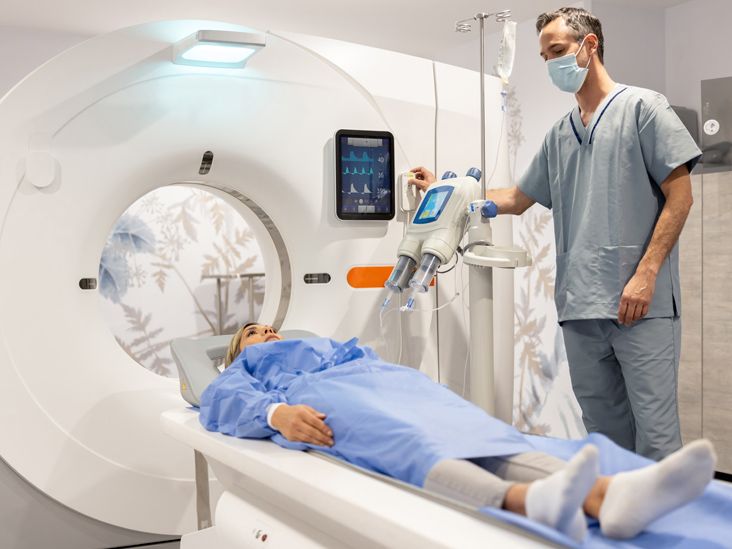
Understanding Radiological Indicators of Crohn’s Disease

Understanding Radiological Indicators of Crohn’s Disease
Crohn's disease is a persistent inflammatory bowel condition that impacts the gastrointestinal tract. Recognized as a form of inflammatory bowel disease (IBD), it manifests through various symptoms and complications. Diagnostic imaging methods such as MRI and CT scans play a crucial role in identifying key signs of this disease, including bowel wall thickening and ulcers. Other imaging modalities like ultrasounds and X-rays may also provide valuable insights.
The Importance of Imaging Studies
While endoscopy—utilizing a miniature camera to inspect the intestines—remains the gold standard for diagnosis, imaging tests complement this process. They help in:
- Eliminating other possible health issues
- Evaluating the severity and extent of the disease
- Monitoring for complications
This article delves into the imaging techniques used to diagnose Crohn's disease and the specific signs that healthcare providers look for during these assessments.
Detection of Crohn’s Disease via MRI
Magnetic Resonance Imaging (MRI) stands out as a premier imaging tool for visualizing inflammation indicative of Crohn’s disease. It effectively highlights complications and disease activity. Key features visible on MRI include:
- Thickened bowel walls
- Strictures (narrowing of the intestines)
- Ulcerations
- Creeping fat (fatty tissue around the intestines)
- Skip lesions (areas of inflammation that are interspersed with healthy tissue)
To give insight into disease progression, doctors often recommend repeated MRIs, which carry no radiation exposure—a significant advantage over CT scans and X-rays.
MRI Technique Types
- MR Enteroclysis: A tube is inserted through the nose leading to the small intestine, where a contrast solution is administered.
- MR Enterography: Patients consume a contrast liquid that fills the bowel, enabling better monitoring of disease activity and treatment effectiveness.
According to a 2018 study, MR Enterography demonstrated superior accuracy compared to ultrasound for various diagnostic metrics related to Crohn’s disease.
Can CT Scans Identify Crohn’s Disease?
Computed Tomography (CT) scans utilize multiple X-rays to produce detailed images of the abdomen. They are instrumental in assessing:
- Activity of the disease
- Extent of inflammation
- Potential complications
Characteristics indicative of Crohn’s disease often observed in CT scans consist of:
- Thickened bowel walls
- Creeping fat
- The “comb sign” indicating swollen blood vessels in the bowel
CT Enterography, a specialized type, can enhance visualization of the small intestines and accurately identifies or rules out Crohn’s disease in over 90% of cases.
Ultrasound's Role in Diagnosing Crohn’s Disease
Ultrasound uses sound waves to generate images without radiation exposure. It can identify Crohn's disease and assess complications affecting other organs, such as the:
- Liver
- Bladder
- Gallbladder
Abdominal ultrasounds provide quick assessments with high accuracy—detecting Crohn’s inflammation approximately 93% of the time. They are particularly effective in visualizing the junction of the large and small intestines.
Are X-rays Useful in Crohn’s Disease Diagnosis?
X-rays have a limited but valuable role in identifying complications of Crohn’s disease, including:
- Ulcers
- Blockages
- Fistulas
Is a Barium Enema Effective for Crohn’s Disease Diagnosis?
A barium enema involves introducing a contrast dye into the rectum to provide clearer X-ray imagery of the bowel. However, this method is less frequently used compared to CT scans or colonoscopies for evaluating changes in the colon.
Conclusion
Imaging studies are essential for doctors to assess the severity of Crohn's disease and identify potential complications. While MRI is often viewed as the most effective imaging method, CT scans and ultrasounds serve as quicker and cost-effective alternatives.
| Imaging Technique | MRI | CT | Ultrasound | X-ray |
|---|---|---|---|---|
| Purpose | Evaluate disease activity, extent, and complications | Evaluate disease activity, extent, and complications | Evaluate disease activity, severity, and complications | Identify complications |
| Cost | High | Moderate | Low | Low |
| Speed | Slow | Fast | Fast | Fast |
| Accuracy | Highly accurate for detecting disease activity | Comparable accuracy to MRI | Less accurate than MRI but still reliable | Limited application |
| Radiation Exposure | No radiation | Radiation exposure | No radiation | Radiation exposure |
